Marked Troponin Elevation with Normal ECG: A Practical Article on Type 2 MI and Myocardial Injury
Case: “Ms. A”
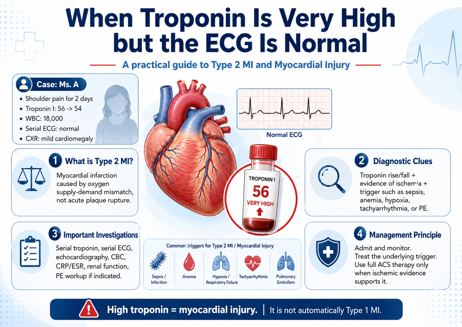
Ms. A presented with shoulder pain for 2 days. Her initial ECG looked normal, with no ST elevation. She did not have active chest pain at the time of reassessment and clinically looked well.
However, her laboratory results were striking:
- Troponin I: 56 → 54
- WBC: 18,000
- BUN: 27
- Creatinine: 0.6
- CXR: normal lungs, mild cardiomegaly
- Serial ECG: completely normal
At first glance, this creates a difficult clinical question:
Is this NSTEMI, Type 2 MI, myocarditis, pulmonary embolism, sepsis-related myocardial injury, or another form of myocardial injury?
The important lesson is this:
A very high troponin confirms myocardial injury, but it does not automatically confirm myocardial infarction.
1. Troponin elevation: what it means and what it does not mean
Troponin is a marker of myocardial cell injury. If troponin is above the 99th percentile upper reference limit, the patient has myocardial injury. But myocardial infarction requires more than just an elevated troponin. It requires myocardial injury plus evidence of acute myocardial ischemia. (American College of Cardiology)
This is why Ms. A’s case is difficult.
Her Troponin I is markedly elevated, but:
- ECG is normal.
- Serial ECG remains normal.
- She has no current chest pain.
- Troponin is 56 → 54, which is persistently high but not clearly rising.
- WBC is high, suggesting inflammation, infection, stress response, or another systemic trigger.
So the correct initial label should not be “definite MI.” A safer and more accurate working diagnosis is:
Marked myocardial injury; rule out NSTEMI, Type 2 MI, myocarditis, pulmonary embolism, and infection-related myocardial injury.
2. What is Type 2 MI?
Type 2 myocardial infarction is MI caused by an imbalance between myocardial oxygen supply and myocardial oxygen demand, not by acute coronary plaque rupture or coronary thrombosis. (American College of Cardiology)
In simple terms:
The heart muscle is injured because it needs more oxygen than it is receiving.
This can happen even without an acute coronary clot.
Common triggers include:
- Sepsis or severe infection
- Severe anemia
- Hypoxia
- Hypotension or shock
- Severe hypertension
- Tachyarrhythmia
- Pulmonary embolism
- Acute heart failure
- Respiratory failure
In Ms. A’s case, the WBC 18,000 is important because infection or systemic inflammation could create a supply–demand mismatch. But Type 2 MI still requires evidence of ischemia. If troponin is elevated without ischemic evidence, the diagnosis is myocardial injury, not Type 2 MI.
3. Type 1 MI vs Type 2 MI vs myocardial injury
| Condition | Main mechanism | Troponin | Ischemia needed? | Example |
| Type 1 MI | Plaque rupture + coronary thrombosis | Elevated, rise/fall | Yes | Classic ACS/NSTEMI/STEMI |
| Type 2 MI | Oxygen supply–demand mismatch | Elevated, rise/fall | Yes | Sepsis with ischemic ECG change and troponin rise |
| Myocardial injury | Myocyte injury without proven ischemia | Elevated | No | Myocarditis, renal failure, sepsis injury, cardiomyopathy |
The AHA scientific statement emphasizes that MI should be reserved for myocardial injury caused by ischemia, whether from atherothrombosis in Type 1 MI or supply–demand mismatch in Type 2 MI. Injury without ischemia should be categorized as myocardial injury rather than MI. (AHA Journals)
4. Pathophysiology of Type 2 MI
The myocardium depends on balance between oxygen supply and oxygen demand.
Myocardial oxygen supply depends on:
- Coronary blood flow
- Arterial oxygen saturation
- Hemoglobin concentration
- Diastolic blood pressure
- Coronary perfusion pressure
- Absence of severe coronary stenosis
Myocardial oxygen demand increases with:
- Tachycardia
- Fever
- Hypertension
- Pain
- Sepsis
- Hyperthyroidism
- Increased wall stress
- Heart failure
Type 2 MI occurs when the supply–demand mismatch becomes severe enough to cause ischemic myocardial necrosis.
For example:
- In sepsis, fever and tachycardia increase oxygen demand, while hypotension and microvascular dysfunction reduce oxygen delivery.
- In anemia, the oxygen-carrying capacity of blood decreases.
- In tachyarrhythmia, oxygen demand rises while coronary filling time falls.
- In pulmonary embolism, right ventricular strain and hypoxia can injure the myocardium.
- In severe hypertension, afterload and wall stress increase myocardial oxygen demand.
5. Diagnostic criteria for Type 2 MI
To diagnose Type 2 MI, the patient must have:
1. Troponin elevation above the 99th percentile
Usually with a rise and/or fall pattern.
2. Evidence of acute myocardial ischemia
At least one of the following:
- Ischemic symptoms
- New ischemic ECG changes
- New pathological Q waves
- Imaging evidence of new regional wall motion abnormality
- Imaging evidence of new loss of viable myocardium
3. A supply–demand mismatch trigger
Examples include sepsis, anemia, hypoxia, shock, tachyarrhythmia, severe hypertension, or pulmonary embolism.
4. No evidence of acute coronary thrombosis
If acute plaque rupture or coronary thrombosis is present, the diagnosis becomes Type 1 MI, not Type 2 MI. (American College of Cardiology)
6. Applying the criteria to Ms. A
Troponin criterion
She clearly has myocardial injury because Troponin I is markedly elevated.
Rise/fall pattern
Troponin I 56 → 54 shows persistent elevation but not a convincing dynamic rise/fall. This may represent late presentation, stable injury, assay variation, or non-acute injury. More serial testing may be needed.
Ischemic evidence
Currently weak:
- No active chest pain
- Serial ECG normal
- No ST elevation
- No ischemic ECG changes
However, shoulder pain can be an anginal equivalent, especially in women or patients with atypical ACS presentations.
Trigger
Possible trigger exists:
- WBC 18,000 suggests infection, inflammation, or stress response.
- Renal failure is unlikely because creatinine is 0.6.
- Pulmonary embolism, myocarditis, and occult infection still need evaluation.
Current conclusion
At this stage, Ms. A does not yet meet a clean diagnosis of Type 2 MI unless ischemic evidence is demonstrated.
The best working diagnosis remains:
Marked myocardial injury, etiology undetermined; rule out NSTEMI, Type 2 MI, myocarditis, pulmonary embolism, and infection-related myocardial injury.
7. Why a normal ECG does not completely exclude MI
A normal ECG reduces the probability of acute coronary occlusion, but it does not fully exclude NSTEMI or myocardial injury. ACS diagnosis depends on clinical presentation, serial ECG, serial troponin, and risk stratification, not a single ECG alone. The 2023 ESC ACS guideline integrates ECG findings, high-sensitivity troponin, clinical assessment, and invasive strategy decisions across the ACS spectrum. (European Society of Cardiology)
Therefore, Ms. A should not be discharged simply because the ECG is normal.
8. Investigation plan
A. Confirm the myocardial injury pattern
Serial Troponin I using the same assay
Do not switch to Troponin T just to “confirm.” Troponin I is already valid. The key is serial change using the same assay.
Purpose:
- Detect rise/fall pattern
- Distinguish acute injury from chronic or stable injury
- Support or weaken the diagnosis of acute MI
CK-MB
Useful when timing is unclear or reinfarction is suspected. It is less sensitive than troponin but can help in selected cases.
B. Look for ischemia
Serial ECG
Already normal in this case, but repeat if:
- Chest pain recurs
- Shoulder pain worsens
- Dyspnea develops
- Hypotension occurs
- Arrhythmia occurs
Echocardiography — the key next test
Echo is very important in this case.
Possible findings:
- Regional wall motion abnormality → supports ischemic MI/NSTEMI
- Global hypokinesia → myocarditis, sepsis cardiomyopathy, cardiomyopathy
- RV dilatation or RV strain → consider pulmonary embolism
- Pericardial effusion → supports myopericarditis
- Low EF or structural heart disease → explains mild cardiomegaly
Coronary CTA or invasive coronary angiography
Consider if:
- Echo shows regional wall motion abnormality
- Recurrent ischemic symptoms occur
- Troponin remains markedly abnormal without another explanation
- High-risk CAD features exist
- Cardiology suspects ACS
C. Search for Type 2 MI triggers and non-ischemic myocardial injury
CBC with differential
Already shows WBC 18,000. Also check hemoglobin because severe anemia can trigger Type 2 MI.
CRP and ESR
Look for systemic inflammation or myocarditis.
Procalcitonin
Useful if bacterial infection or sepsis is suspected.
Blood culture
If fever, chills, hypotension, or suspected sepsis.
Urinalysis and urine culture
To look for occult UTI.
Electrolytes, magnesium, calcium
Arrhythmia and myocardial stress can be worsened by electrolyte disorders.
TSH/free T4
Hyperthyroidism can cause tachycardia and demand ischemia.
ABG/VBG and lactate
If hypoxia, sepsis, shock, or poor perfusion is suspected.
D-dimer or CTPA
If pulmonary embolism is clinically suspected, especially with dyspnea, tachycardia, hypoxia, pleuritic pain, hemoptysis, DVT signs, recent immobilization, cancer, pregnancy, or estrogen use.
Cardiac MRI
Useful when myocarditis is suspected, and echo/coronary evaluation is inconclusive.
9. Management principles
First principle: admit and monitor
Ms. A should be admitted or observed in a monitored setting because Troponin I 56 is markedly abnormal.
Recommended:
- Telemetry
- Serial vital signs
- Serial ECG if symptoms recur
- Repeat Troponin I
- Echocardiography
- Cardiology consultation
- Search for infection, PE, myocarditis, arrhythmia, anemia, and hypoxia
10. Drug management in this difficult situation
Because ACS is not fully excluded, some ACS-protective treatment may be reasonable if there is no contraindication.
Reasonable while evaluating
Aspirin 160–325 mg chew stat, then 81 mg PO daily. Reasonable if bleeding risk is low and ACS remains possible.
Atorvastatin 40–80 mg PO stat, then dailyReasonable because ACS remains in the differential and vascular risk may be present.
Hold unless ACS becomes more likely
Do not automatically start full NSTEMI therapy with dual antiplatelet therapy and anticoagulation unless ischemic evidence appears.
Be cautious with:
- Clopidogrel
- Ticagrelor
- Enoxaparin
- Unfractionated heparin
These are most useful when coronary thrombosis or true ACS is likely. In Type 2 MI or non-ischemic myocardial injury, routine aggressive antithrombotic therapy may expose the patient to bleeding risk without treating the underlying cause.
When to give full NSTEMI treatment
Escalate to full ACS/NSTEMI management if any of the following appears:
- Recurrent ischemic chest pain or anginal equivalent
- New ischemic ECG changes
- New regional wall motion abnormality on echo
- Hemodynamic instability
- High clinical suspicion of coronary ACS
- Cardiology recommends invasive ACS strategy
11. Management if Type 2 MI is confirmed
The main treatment of Type 2 MI is to correct the supply–demand mismatch.
Examples:
If infection/sepsis is the trigger
- Culture before antibiotics if possible
- Start appropriate antibiotics early
- Give fluids if hypovolemic
- Use vasopressors if septic shock
- Control fever
- Monitor lactate and organ perfusion
If anemia is the trigger
- Identify bleeding source
- Transfuse if severe anemia, active ischemia, or hemodynamic compromise
- Treat underlying cause
If hypoxia is the trigger
- Oxygen if hypoxemic
- Treat pneumonia, PE, COPD/asthma exacerbation, or respiratory failure
If tachyarrhythmia is the trigger
- Rate control or rhythm control
- Correct electrolytes
- Treat underlying infection, thyrotoxicosis, or heart failure
If pulmonary embolism is the trigger
- Anticoagulation if confirmed or strongly suspected
- Risk stratify for thrombolysis or intervention if massive/submassive PE
If severe hypertension is the trigger
- Controlled BP reduction
- Avoid overly rapid hypotension
- Treat end-organ injury
12. Common pitfalls in cases like Ms. A
Pitfall 1: “Troponin high = MI”
Wrong. Troponin high = myocardial injury. MI requires ischemia.
Pitfall 2: “Normal ECG = safe discharge”
Wrong. Normal ECG does not fully exclude NSTEMI, myocarditis, PE-related myocardial injury, or other dangerous causes.
Pitfall 3: “Type 2 MI = any sick patient with troponin”
Wrong. Type 2 MI still needs evidence of myocardial ischemia.
Pitfall 4: “Troponin T is needed to confirm Troponin I”
Wrong. Use the same troponin assay serially. Switching assays can confuse interpretation.
Pitfall 5: “Start full DAPT and anticoagulation for every high troponin”
Not always. If the cause is myocarditis, sepsis-related injury, PE, or non-ischemic myocardial injury, treatment should target the underlying cause.
13. Practical diagnostic wording for this case
The best clinical wording is:
Markedly elevated Troponin I / myocardial injury with normal serial ECG and no active chest pain; rule out NSTEMI, Type 2 MI, myocarditis, pulmonary embolism, and infection-related myocardial injury.
This wording is safer than immediately diagnosing NSTEMI.
14. Practical management summary for Ms. A
- Admit to telemetry or monitored unit
- Cardiology consult
- Repeat Troponin I using same assay
- Urgent echocardiography
- Continue serial ECG if symptoms recur
- Give aspirin and high-intensity statin if no contraindication
- Do not automatically give full DAPT/enoxaparin unless ischemic evidence appears
- Investigate leukocytosis
- CBC differential
- CRP/ESR
- Procalcitonin
- UA/urine culture
- Blood culture if febrile
- Assess for PE
- SpO₂, HR, RR
- DVT signs
- D-dimer/CTPA if clinically suspicious
- Consider myocarditis
- Viral prodrome
- CRP/ESR
- Echo Heart
- Cardiac MRI if needed
Final teaching point
This case is difficult because the troponin is very high, but the ischemic evidence is weak.
The correct thinking is not:
“Troponin I 56 = definite MI.”
The correct thinking is:
“Troponin I 56 = serious myocardial injury. Now I must determine whether it is Type 1 MI, Type 2 MI, myocarditis, PE-related injury, sepsis-related injury, or another cause.”
Comments
No comments yet. Be the first to share your thoughts.
Sign in to comment