Ottawa Ankle Rules: When to Order X-rays in Ankle Injuries
Introduction
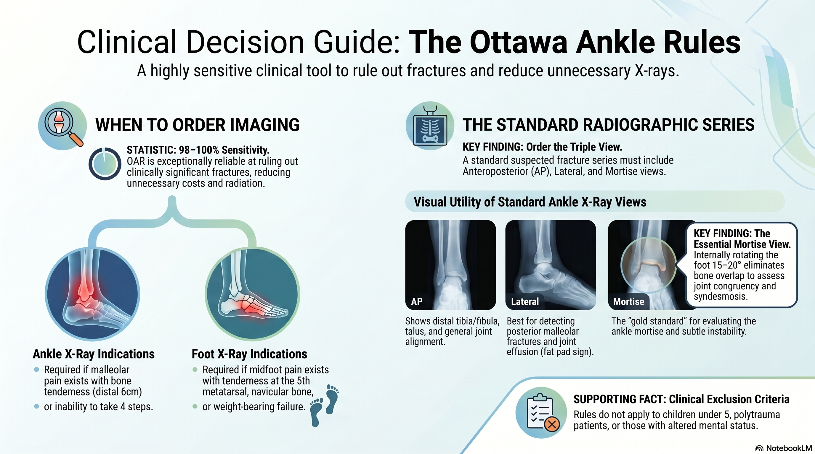
Ankle injuries are one of the most common presentations in emergency and outpatient settings. However, not all patients require radiographic imaging. The Ottawa Ankle Rules (OAR) are a validated clinical decision tool designed to reduce unnecessary X-rays while maintaining high sensitivity for detecting clinically significant fractures.
Ottawa Ankle Rules (OAR)
ⓒ Original publisher. Displayed via hotlinking for educational fair use; fallback to archived copy if unavailable.
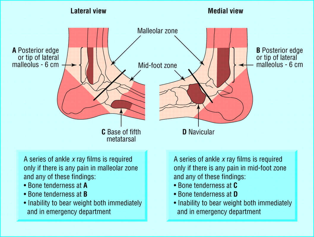
Indications for Ankle X-ray
An ankle X-ray series is indicated only if there is pain in the malleolar zone AND at least one of the following:
- Bone tenderness at the posterior edge or tip of the lateral malleolus (distal 6 cm), OR
- Bone tenderness at the posterior edge or tip of the medial malleolus (distal 6 cm), OR
- Inability to bear weight both immediately after injury and in the emergency department (unable to take 4 steps)
Indications for Foot X-ray (for completeness)
If there is pain in the midfoot zone AND at least one of the following:
- Tenderness at the base of the 5th metatarsal, OR
- Tenderness at the navicular bone, OR
- Inability to bear weight (same criteria)
Clinical Importance
- Sensitivity: ~98–100% (very good at ruling out fractures)
- Helps reduce unnecessary radiation and cost
- Widely recommended in emergency medicine guidelines
Standard Ankle Radiographic Views
When Ottawa Ankle Rules indicate imaging → order:
1. Anteroposterior (AP) View
- Foot in neutral position
- Shows:
- Distal tibia and fibula
- Talus
- Joint alignment
- Limitation: Overlap of tibia and fibula may obscure syndesmosis
2. Lateral View
- Foot positioned laterally
- Shows:
- Talus, calcaneus
- Posterior malleolus
- Joint effusion (fat pad sign)
- Important for detecting:
- Posterior malleolar fractures
- Displacement
3. Mortise View (Most Important)
- Foot internally rotated 15–20 degrees
- Eliminates overlap between tibia and fibula
- Provides clear visualization of:
- Ankle mortise (tibiotalar joint)
- Syndesmosis
- Key for:
- Detecting subtle fractures
- Assessing joint congruency and instability
Summary
- Use Ottawa Ankle Rules → decide whether to send X-ray
- If indicated → order 3 standard views: AP + Lateral + Mortise
- Mortise view = best for ankle joint assessment
Common OSCE / Exam Question Tip
If asked:👉 “What imaging do you order for a suspected ankle fracture?” Answer: “Ankle X-ray including AP, lateral, and mortise views.”
Clinical Caution ⚠️
- Ottawa rules cannot be used in:
- Children <5 years
- Altered mental status
- Multiple trauma
- Always consider clinical judgment beyond rules
Comments
No comments yet. Be the first to share your thoughts.
Sign in to comment