Dyshidrotic Eczema (Pompholyx): Review, Diagnosis, and Management
Dyshidrotic Eczema (Pompholyx)
Spot diagnosis
- Intensely itchy
- Deep, clear vesicles
- Palms, sides of fingers, soles
- “Tapioca-like” appearance
Triggers
- Stress, sweating
- Nickel/cobalt
- Irritants, atopy
Diagnosis
- Clinical
- KOH only if tinea suspected
Management (OPD)
- High-potency topical steroid
- Clobetasol 0.05% ointment BID × 7–14 days
- Emollients frequently
- Cold compress
Severe flare
- Prednisone 0.5–1 mg/kg/day PO × 5–7 days (short course)
Refractory
- Tacrolimus ointment
- Phototherapy
- Treat hyperhidrosis
Avoid
- Routine antibiotics
- Long-term systemic steroids
Key pearl
- Not sweat duct obstruction; eczema with high recurrence.
ⓒ Original publisher. Displayed via hotlinking for educational fair use; fallback to archived copy if unavailable.
ⓒ Original publisher. Displayed via hotlinking for educational fair use; fallback to archived copy if unavailable.
Dyshidrosis (Dyshidrotic eczema / Pompholyx) — doctor-level review and management
1) Definition and clinical phenotype
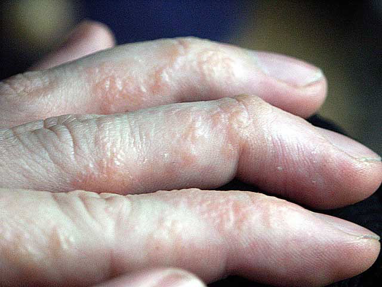
Dyshidrosis is a recurrent vesicular eczema affecting palms, lateral fingers, and soles, characterized by intensely pruritic, deep-seated vesicles that look like “tapioca pearls,” followed by desquamation, fissuring, and lichenification in chronic disease. It is best conceptualized as a subtype of chronic hand eczema (CHE) (often “recurrent vesicular hand eczema”). (arts.units.it)
2) Epidemiology and associations (what matters clinically)
- Hand eczema is common; dyshidrosis represents a recognizable vesicular phenotype within it. (arts.units.it)
- Strong associations:
- Atopy (personal/family history of atopic dermatitis, allergic rhinitis, asthma)
- Irritant exposure (wet work, detergents, solvents, frequent handwashing)
- Allergic contact dermatitis (notably nickel/cobalt/chromate)
- Hyperhidrosis and psychological stress as flare triggers (arts.units.it)
3) Pathophysiology (board-style, but clinically useful)
Think “eczema biology,” not sweat gland obstruction:
- Skin barrier dysfunction (reduced barrier lipids/filaggrin-related pathways in some patients) → increased irritant penetration and immune activation.
- Inflammation with spongiosis in epidermis → vesicle formation (the “tapioca” vesicles are essentially intraepidermal spongiotic vesicles).
- Triggers (irritants/allergens/stress/sweat) amplify immune signaling and barrier breakdown.The ESCD hand eczema guideline frames dyshidrosis within broader hand eczema evaluation (irritant + allergic + atopic contributors often overlap). (arts.units.it)
4) Clinical features and staging (how to “read” the rash)
Distribution
- Palms, lateral fingers; sometimes soles/toes.
- Dorsal hand involvement can occur but should raise concern for contact dermatitis pattern.
Morphology over time
- Prodrome: itch/burning/tingling hours–days before eruption.
- Acute vesicular phase: deep, tense vesicles; intense pruritus, sometimes pain.
- Subacute: vesicles dry → scaling/peeling.
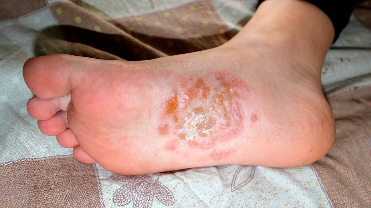
- Chronic: fissures, thickening/lichenification; secondary infection risk increases.
Clues that suggest a “driver”
- Dominant palmar/side-of-finger vesicles with recurrent flares: classic dyshidrosis phenotype.
- Clear exposure pattern (new gloves, sanitizer, metal contact): consider allergic/irritant contact dermatitis coexisting.
- Unilateral involvement, annular scale, moccasin foot: consider tinea.
5) Differential diagnosis (and how to rule out efficiently)
- Allergic contact dermatitis (ACD)
- Often dorsal hands/wrists, sharp borders, exposure correlation.
- Confirm with patch testing. (arts.units.it)
- Irritant contact dermatitis (ICD)
- “Wet work” history; burning > itch; chronic dryness/fissures.
- Diagnosis is clinical; prevention is central. (arts.units.it)
- Tinea manuum / pedis (dermatophyte)
- Often unilateral hand (“two feet–one hand” pattern).
- Do KOH and/or fungal culture. (arts.units.it)
- Palmoplantar pustulosis / psoriasis
- Sterile pustules, erythema, nail pitting/onycholysis; smoker association.
- Scabies
- Web spaces, burrows, nocturnal itch, household contacts.
- Herpetic whitlow (important “don’t miss”)
- Grouped vesicles, marked pain, possible systemic symptoms; consider if healthcare worker or HSV history.
6) Diagnostic work-up (what a dermatologist expects)
Dyshidrosis is usually clinical, but recurrent/severe disease benefits from structured evaluation.
Minimum evaluation in recurrent hand/foot vesicles
- Exposure history: wet work, soaps/sanitizers, gloves (rubber/latex), occupational chemicals, metals.
- Atopy history.
- Hyperhidrosis assessment.
- Fungal rule-out: KOH scraping if any scale/unilateral pattern/foot involvement. (arts.units.it)
When to order patch testing
- Chronic/recurrent hand eczema, severe disease, or suspicion of ACD (new products, occupational exposure, distribution suggests contact).ESCD/S2k guideline emphasizes patch testing as a key tool when ACD is possible. (arts.units.it)
Severity documentation (useful for follow-up and referrals)
- HECSI (Hand Eczema Severity Index) or a simpler “photographic + functional impairment” documentation is often used in studies and specialty care. (Dermatology Times)
7) Management (stepwise, practical, guideline-aligned)
A) Setting: OPD vs IPD
- Most patients: OPD (even severe flares).
- Consider IPD only if: extensive secondary infection/cellulitis requiring IV antibiotics, severe functional impairment with inability to care for self, or diagnostic uncertainty with systemic illness.
(Hand eczema guidelines are outpatient-focused; escalation is typically via specialty care rather than admission.) (arts.units.it)
B) Core principle: treat three targets every time
- Inflammation control (anti-inflammatory therapy)
- Barrier repair (emollients + protection)
- Trigger control (irritant reduction, sweat control, allergy identification) (arts.units.it)
C) Patient Problem List (typical for dyshidrosis)
- Acute vesicular hand/foot eczema flare (dyshidrosis phenotype)
- Skin barrier failure (xerosis, fissuring, pain)
- Pruritus affecting sleep/function
- Possible triggers: irritant exposure / hyperhidrosis / suspected contact allergy
- Monitor for secondary infection
Problem 1: Dyshidrosis flare (vesicular hand/foot eczema)
Definitive treatment (first-line)
High-potency topical corticosteroid (palms/soles need higher potency; ointment penetrates better)
- Clobetasol propionate 0.05% (thin layer) BID topical for 7–14 daysor
- Betamethasone dipropionate 0.05% (thin layer) BID topical for 7–14 days
Key technique: apply after a short soak, then ointment, then consider short-term occlusion (cotton glove) for a few nights if very thick skin—avoid prolonged occlusion if maceration/infection risk.
This aligns with hand eczema guideline frameworks emphasizing topical corticosteroids as mainstay anti-inflammatory therapy. (arts.units.it)
Supportive treatment (during acute vesicular stage)
- Wet compresses (to dry vesicles and reduce itch):
- Aluminum acetate (Burow solution) compress 10–15 min, 2–4×/day for several days(common dermatology practice; used as symptomatic control in vesicular hand eczema) (American Academy of Dermatology)
- If itch disrupts sleep:
- Hydroxyzine (25 mg) 1×1 PO HS PRNor non-sedating daytime option: cetirizine (10 mg) 1×1 PO daily.
Escalation for severe flare (short course)
- Prednisone 0.5 mg/kg/day PO for 5–7 days, then stop or taper if longer course is used.Use only for acute rescue; relapse can occur if triggers not controlled.
Guidelines generally reserve systemic steroids for short-term rescue rather than maintenance in chronic hand eczema. (arts.units.it)
Why not antibiotics routinely
Antibiotics are not indicated unless there is evidence of infection (oozing pus, honey crusting, increasing erythema/warmth, lymphangitis, fever). This is consistent with rational management of eczema and chronic hand eczema guidance emphasizing targeted therapy. (arts.units.it)
Problem 2: Barrier failure (dryness, scaling, fissures)
Definitive treatment (barrier repair)
- Petrolatum-based ointment or fragrance-free thick emollient at least 4–6×/day, always after handwashing.
- Night regimen: emollient + cotton gloves.
Supportive / protective measures (reduce irritant load)
- Replace soap-and-water with alcohol-based hand rub when hands are not visibly soiled (less irritating than repeated soap-and-water in many cases).
- “Glove protocol”:
- Wet work: nitrile/vinyl gloves
- Always use cotton liner and limit continuous wear; remove when sweating starts
- Avoid hot water, harsh detergents, fragranced products.
Hand eczema guidelines emphasize emollients and irritant avoidance as foundational therapy. (arts.units.it)
Problem 3: Pruritus and pain (functional impairment)
Definitive treatment
- Control inflammation (topical steroid is the pruritus treatment).
Supportive treatment
- Sedating antihistamine at night if sleep is impaired (as above).
- For fissure pain: thick ointment + occlusion; consider cyanoacrylate skin protectant for single fissures (clinical practice).
Problem 4: Trigger control (hyperhidrosis, allergy, occupational factors)
1) Hyperhidrosis-related flares
- Aluminum chloride hexahydrate 20% apply nightly to palms/soles; reduce frequency as tolerated.
- Consider tap-water iontophoresis or botulinum toxin via dermatology for refractory cases (specialist-level). (Medscape)
2) Suspected allergic contact dermatitis contributor
- Arrange patch testing for recurrent/chronic disease, especially with occupational exposure or poor response to standard therapy. (arts.units.it)
3) Fungal trigger (id reaction)
- If tinea pedis is present, treat it; it can perpetuate hand eczema in some patients. Hand eczema guidelines include ruling out and addressing infection triggers. (arts.units.it)
8) Refractory disease (dermatology escalation path)
If inadequate control after appropriate potency topical therapy + trigger control, consider escalation consistent with CHE guidance.
Steroid-sparing topical maintenance
- Tacrolimus 0.1% ointment BID topical (often as maintenance or for frequent relapses). (arts.units.it)
Phototherapy
- Narrowband UVB or PUVA can be effective for chronic/refractory hand eczema. (arts.units.it)
Systemic options (specialist-driven; consider local availability)
- Alitretinoin 30 mg 1×1 PO daily for 12–24 weeks for severe chronic hand eczema refractory to topical steroids (commonly used in Europe/Canada; requires strict pregnancy prevention, lipid/LFT monitoring). (arts.units.it)
- Immunosuppressants (off-label in many settings): cyclosporine, methotrexate, azathioprine, mycophenolate—choice depends on phenotype, comorbidities, monitoring capacity. (arts.units.it)
- Dupilumab: evidence supports benefit in severe chronic hand eczema subtypes including recurrent vesicular hand eczema; dosing in trials typically 600 mg loading then 300 mg every 2 weeks. (OUP Academic)
9) Monitoring, complications, and safety checks
Monitor treatment response
- Symptom control (itch, pain)
- Function (hand use, work ability)
- Skin integrity (fissures)
- Frequency of relapses
- Consider severity scoring (HECSI) in recurrent disease or specialty referral. (Dermatology Times)
Watch for infection
- Increasing erythema, warmth, tenderness, purulence, fever.
- If suspected, culture if significant drainage; treat appropriately.
Steroid safety (topical)
- High potency on palms is appropriate but limit continuous use:
- Typical burst 1–2 weeks, then taper frequency or switch to maintenance regimen.
- Avoid chronic daily superpotent use without breaks (atrophy risk is lower on palms but still relevant).
10) Follow-up plan (what to write in the chart)
- Follow up in 2–4 weeks after initiating high-potency topical steroid burst.
- If improved: step down to intermittent steroid (weekend therapy) or calcineurin inhibitor maintenance + strict barrier care.
- If not improved:
- confirm diagnosis (KOH, patch testing),
- reassess exposures and adherence,
- consider phototherapy/systemic therapy referral. (arts.units.it)
11) Red flags (when to escalate urgently)
- Severe pain with grouped vesicles suggesting herpes (herpetic whitlow)
- Rapidly spreading erythema, fever, systemic symptoms (cellulitis)
- Widespread blistering beyond typical distribution
- Immunosuppressed patient with worsening lesions
Comments
No comments yet. Be the first to share your thoughts.
Sign in to comment